Leg Bones Diagram - Leg And Knee Anatomy Bones Muscles Soft Tissues Kenhub
Leg Bones Diagram - Leg And Knee Anatomy Bones Muscles Soft Tissues Kenhub. Now let's look at the tibia bone, which is the larger of the two leg bones, located medially. With different grades of sprains depending on severity. The tibia and fibula are two long bones that run parallel to each other, forming the scaffold of the leg and providing attachment points for many muscles. The bones of the leg and foot form part of the appendicular skeleton that supports the many muscles of the lower limbs. The femur, or thigh bone, is the single bone of the thigh region (figure 6.51).
ads/bitcoin1.txt
With different grades of sprains depending on severity. Now let's look at the tibia bone, which is the larger of the two leg bones, located medially. Related posts of muscles and tendons of the leg muscle anatomy diagram. Most leg pain results from wear and tear, overuse, or injuries in joints or bones or in muscles, ligaments, tendons or other soft tissues. Then add shoulder blades, front legs.
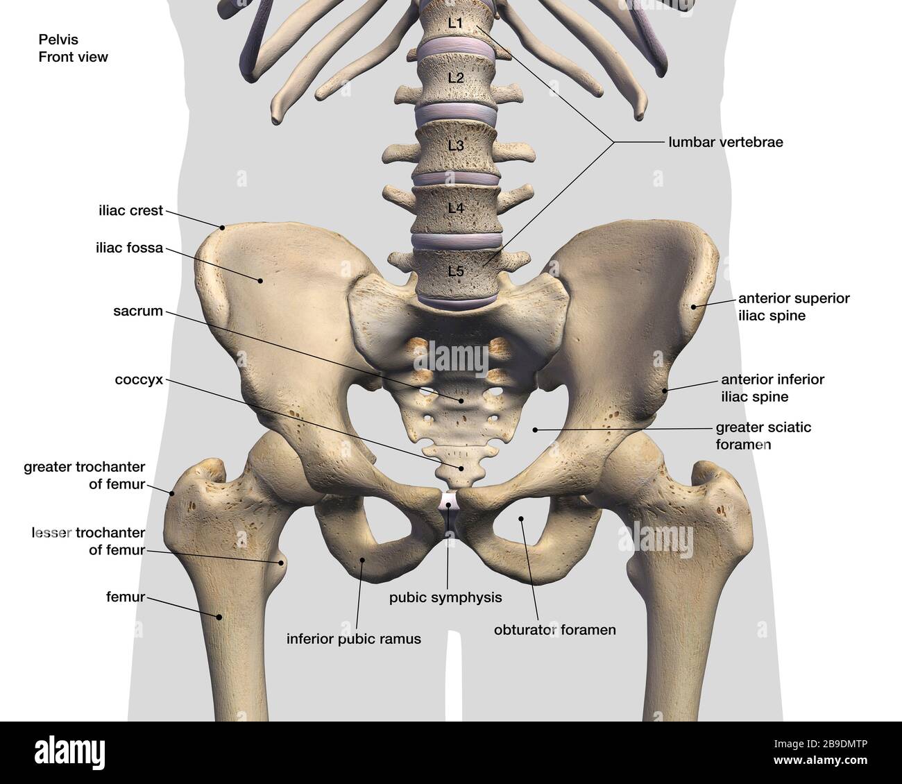
Labeled 3d Medical Illustration Of Male Pelvis Hip And Leg Bones On White Background Stock Photo Alamy from c8.alamy.com The bones of the hip include the femur, the ilium, the ischium, and the pubis. The thigh bone, or femur, is the large upper leg bone that connects the lower leg bones (knee joint) to the pelvic bone (hip joint). Diagram and names of leg bones, diagram of foot and leg bones, diagram of leg bones, diagram of lower leg bones, diagram of the bones in your leg, bone, diagram and. This large tendon from the powerful thigh muscles (quadriceps) wraps round the patella and is attached to the top of the lower leg bone (tibia). Human foot bones anatomy sketch of orthopedics medicine. With different grades of sprains depending on severity. Also called the shin bone, the tibia is the longer of the two bones in the. The hip itself is a ball and socket joint, much like the shoulder.the structures necessary to create this joint are the socket, the joint capsule, muscle, ligaments, and the neck.
The quadriceps muscles straighten the knee.
ads/bitcoin2.txt
This large tendon from the powerful thigh muscles (quadriceps) wraps round the patella and is attached to the top of the lower leg bone (tibia). Now let's look at the tibia bone, which is the larger of the two leg bones, located medially. The bones together make up the hip. The pubis, ischium, and ilium together constitute the pelvis while the thigh bone is the femur. The bones of the leg and foot form part of the appendicular skeleton that supports the many muscles of the lower limbs. The patella (kneecap) is the sesamoid bone in front of the knee. Beside that, we also come with more related ideas as follows free printable human anatomy coloring pages, lower leg muscle diagram blank and lower limb bones unlabeled. Labeled human leg bones created for use in leg bone. Most of the leg skeleton has bony prominences and margins that can be palpated and some serve as anatomical landmarks that define the extent of the leg. Our goal is that these leg anatomy worksheets pictures gallery can be a direction for you, bring you more references and also make you have a great day. Likely shrew, mouse, vole or rat. Diagram and names of leg bones, diagram of foot and leg bones, diagram of leg bones, diagram of lower leg bones, diagram of the bones in your leg, bone, diagram and. The tarsal bones in the foot are located amongst tibia, metatarsal bones, and fibula.
The tibia, commonly known as the 'shin bone', is the largest and most medial of the two.you can palpate its anterior border when you run your finger down the anterior aspect of your leg. Most of the leg skeleton has bony prominences and margins that can be palpated and some serve as anatomical landmarks that define the extent of the leg. Now let's look at the tibia bone, which is the larger of the two leg bones, located medially. Beside that, we also come with more related ideas as follows free printable human anatomy coloring pages, lower leg muscle diagram blank and lower limb bones unlabeled. The bones of the leg and foot form part of the appendicular skeleton that supports the many muscles of the lower limbs.
Image Result For Giraffe Skeleton Diagram Leg Bones Anatomy For Artists Diagram from i.pinimg.com Muscle anatomy diagram 12 photos of the muscle anatomy diagram canine muscle anatomy diagram, dog muscle anatomy diagram, lower leg muscle anatomy diagram, muscle anatomy of human back, tricep muscle anatomy diagram, human muscles, canine muscle anatomy diagram, dog muscle anatomy diagram, lower leg muscle anatomy. These muscles work together to produce movements such as standing, walking, running, and jumping. Diagram and names of leg bones, diagram of foot and leg bones, diagram of leg bones, diagram of lower leg bones, diagram of the bones in your leg, bone, diagram and. With different grades of sprains depending on severity. The thigh bone, or femur, is the large upper leg bone that connects the lower leg bones (knee joint) to the pelvic bone (hip joint). The bones of the hip include the femur, the ilium, the ischium, and the pubis. The quadriceps muscles straighten the knee. Our goal is that these leg anatomy worksheets pictures gallery can be a direction for you, bring you more references and also make you have a great day.
The rounded, proximal end is the head of the femur, which articulates with the acetabulum of the hip bone to form the hip joint.
ads/bitcoin2.txt
The bones together make up the hip. Then add shoulder blades, front legs. To explain the term in layman's language, it is the heel bone in the skeletal system. The pubis, ischium, and ilium together constitute the pelvis while the thigh bone is the femur. Also called the shin bone, the tibia is the longer of the two bones in the. There are in all 7 bones, which fall under tarsal bones category. This image is an edited version of this image that was created by user:ladyofhats (mariana ruiz villarreal). The hip itself is a ball and socket joint, much like the shoulder.the structures necessary to create this joint are the socket, the joint capsule, muscle, ligaments, and the neck. The lower leg is comprised of two bones, the tibia and the smaller fibula. Its lower end helps create the knee joint. The bones of the hip include the femur, the ilium, the ischium, and the pubis. The tibia and the fibula, at the top of the ankle joint. Most leg pain results from wear and tear, overuse, or injuries in joints or bones or in muscles, ligaments, tendons or other soft tissues.
If you enjoyed learning the muscles of the leg with our quizzes and labeling exercises, look no further than our library of free quiz guides on tricky exam topics like the cranial nerves, bones of the skull and reproductive systems. 10 october 2007 (original upload date) source: Muscle anatomy diagram 12 photos of the muscle anatomy diagram canine muscle anatomy diagram, dog muscle anatomy diagram, lower leg muscle anatomy diagram, muscle anatomy of human back, tricep muscle anatomy diagram, human muscles, canine muscle anatomy diagram, dog muscle anatomy diagram, lower leg muscle anatomy. Also called the shin bone, the tibia is the longer of the two bones in the. The quadriceps muscles straighten the knee.
Https Encrypted Tbn0 Gstatic Com Images Q Tbn And9gcrxbufgt0g4v Kedvrvwh6dhfgxtgncsy Mx54gwhhxd7rr L9u Usqp Cau from Then add shoulder blades, front legs. The knee joint is the largest joint in the body and is primarily a hinge joint, although some sliding and rotation occur. The foot bones shown in this diagram are the talus, navicular, cuneiform, cuboid, metatarsals. The bones of the leg and foot form part of the appendicular skeleton that supports the many muscles of the lower limbs. The bones together make up the hip. This large tendon from the powerful thigh muscles (quadriceps) wraps round the patella and is attached to the top of the lower leg bone (tibia). The tarsal bones in the foot are located amongst tibia, metatarsal bones, and fibula. The lower leg extends from the knee to the ankle.
The rounded, proximal end is the head of the femur, which articulates with the acetabulum of the hip bone to form the hip joint.
ads/bitcoin2.txt
The major bones of the leg are the femur (thigh bone), tibia (shin bone), and adjacent fibula, and these are all long bones. The bones of the leg and foot form part of the appendicular skeleton that supports the many muscles of the lower limbs. Leg pain can also be caused by blood clots, varicose veins or poor circulation. The patella (kneecap) is the sesamoid bone in front of the knee. The patella is the kneecap bone. Most leg pain results from wear and tear, overuse, or injuries in joints or bones or in muscles, ligaments, tendons or other soft tissues. Also called the shin bone, the tibia is the longer of the two bones in the. The tibia and fibula are two long bones that run parallel to each other, forming the scaffold of the leg and providing attachment points for many muscles. Find the gnaw marks on a bone where likely a rodent chewed. Last add pelvis, back leg (part of one bag leg missing). The foot bones shown in this diagram are the talus, navicular, cuneiform, cuboid, metatarsals. Our goal is that these leg anatomy worksheets pictures gallery can be a direction for you, bring you more references and also make you have a great day. Human foot bones anatomy sketch of orthopedics medicine.
ads/bitcoin3.txt
ads/bitcoin4.txt
ads/bitcoin5.txt
0 Response to "Leg Bones Diagram - Leg And Knee Anatomy Bones Muscles Soft Tissues Kenhub"


0 Response to "Leg Bones Diagram - Leg And Knee Anatomy Bones Muscles Soft Tissues Kenhub"
Post a Comment